This website uses cookies so that we can provide you with the best user experience possible. Cookie information is stored in your browser and performs functions such as recognising you when you return to our website and helping our team to understand which sections of the website you find most interesting and useful. More information in our Privacy Policy
Scanning Electron Microscopes
Supervisors: Mauro Mazzocchi
Electron microscopy allows the observation of objects with magnifications and resolution over 1000 times higher than optical microscopy, acting through an electron beam that interacts with the sample.
The matter of the sample scatters electrons and X-rays with peculiar characteristics, which are collected by suitable detectors to provide topographic information (SE), useful for distinguishing different phases (BSE) and for local chemical analysis (RX) by going back to concentrations and distributions of single elements or phases in the sample.
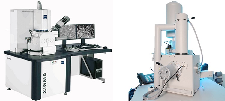
Field emission electron scanning microscope FE-SEM, with EDS and STEM probe, ZEISS SIGMA, Carl Zeiss Microscopy GmbH
- W-ZrO2 source with Schottky effect and hot cathode gun;
- Gemini ™ electro-optical column (ZEISS) with no electromagnetic crossover lenses, with beam booster (+ 8kV) on the liner, for low electronic acceleration voltages (EHT, down to 100 eV) observations and High-Current Mode on probe current, with 100 eV EHT at 30KeV;
- High resolution Imaging (~ 0.1nm);
- Detector Everhart-Thornley for detection of secondary electrons, SE2;
- In-Lens coaxial annular detector for low energy electrons, SE1;
- ASB-CBZD 4-sector ring detector (Angular Selective BSE) with topo-phase effect for backscattered secondary electrons (> 50 eV);
- Energy Dispersive Spectroscopy micro-analysis system (EDS) device Oxford Energy X-Act, with 10 mm² active area SDD detector, interfaced by INCA software for compositional, point and area analysis;
- Resolution up to 125 eV (measured according to ISO 15632: 2002);
- STEM (Scanning transmission electron microscopy) probe is installed (Zeiss) with 12X specimen stage carousel with multiple Si diodes detectors, for signals in bright field (BF) and dark field (DF), both from the annular detector DF (ADF), and from the segments prepared for the high degrees angles (HAADF).
Environmental variable pressure electron scanning microscope ESEMTM Quanta 200, FEI (THERMO FISHER SCIENTIFIC Inc.)
A tungsten thermoionic effect gun supplies a focused electron beam (EHT from 100eV to 30 keV) able to operate imaging in a variable pressure (vacuum with in-room atmosphere of H2O molecules) through 3 working modes:
- High Vacuum (P <10-4 Torr), for conductive and/or metallized specimen;
- Low Vacuum (P <1.5 Torr), allows the analysis of non-dehydrated and -above all- non-conductive specimen, such as organic materials in a natural state without the need for metallization to make them conductive;
- Wet environment, ESEM™, (P> 20 Torr), for the observation of high water content specimen, wet or even preserved in water specimen. The ESEM mode does not require sample preparation and above all it is not invasive on materials because it provides to maintain the sample unaltered in its structural characteristics, allowing a characterization of non-conductive and even wet specimen in their natural state. A multi-purpose Centaur (KE Developments Ltd.) scintillator detector is installed for Cathodoluminescence (300-650 nm) and backscattered electrons (CL / BSD) with high sensitivity even at low energies (from 500 eV) particularly useful for LowVac and ESEM analyses. The main features include:
- Everhart-Thornley Detector for Secondary Electrons SE2 (SED) (HiVac);
- Detector secondary electrons (LFD) (LoVac);
- Secondary electron detector (GSED) (ESEM ™);
- Annular 2-Si-diodes segments detector for backscattered electrons (BSE, > 50 eV) (Hi & LoVac);
- Simultaneous secondary and backscattered electron detectors (GBSD) (ESEM ™);
- Backscattered electron detector (GAD) optimized for EDS analysis (LoVac mode);
- Cooling table-sample holder (Peltier) for wet biological specimen (-25 / + 55 ° C) and in water analysis;
- Cartesian eucentric stage with 4 motorized axes with 7X sample holder carousel; a single specimen holder is available.
Specific equipment is used for samples’ pretreatments, such as:
- Sputter coater: QUORUM mod. Q150T- conductive films (Au, Pt, Pt / Pd) for high resolution imaging, combined with carbon evaporator;
- Low pressure plasma cleaner: Gambetti Kenologia mod. Colibri with double gas line and mass flow controller. High frequency RF generator working at 13.56 MHz and up to 200W.
The microscopy laboratory is also supported by the Cutting and Lapping Laboratory, which has a range of instruments necessary for the optical lapping of planar surfaces. Polished sections can also be prepared by incorporation in cold or hot resin. In particular, the following features are available:
- Automatic lapping machine Struers Tegramin-25 with sample holder up to 6 slots, magnetic fixing of the abrasive discs and automatic dosage of the diamond suspensions;
- Manual lapping machines;
- Metallographic cutting machines with micrometric adjustment;
- Hot and cold embedding machine.

